Showing posts with label aldosterone. Show all posts
Sunday, June 23, 2013

Adrenal Glands

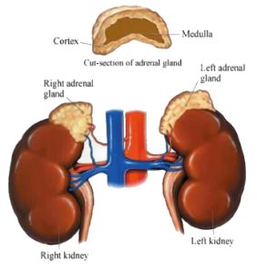
adrenal-glandsAnatomy of the adrenal glands:
Adrenal glands, which are also called suprarenal glands, are small, triangular glands located on top of both kidneys. An adrenal gland is made of two parts: the outer region is called the adrenal cortex and the inner region is called the adrenal medulla.

Function of the adrenal glands:
The adrenal glands work interactively with the hypothalamus and pituitary gland in the following process:
  • the hypothalamus produces corticotropin-releasing hormones, which stimulate the pituitary gland.
  • the pituitary gland, in turn, produces corticotropin hormones, which stimulate the adrenal glands to produce corticosteroid hormones.
Both parts of the adrenal glands -- the adrenal cortex and the adrenal medulla -- perform very separate functions.

What is the adrenal cortex?
The adrenal cortex, the outer portion of the adrenal gland, secretes hormones that have an effect on the body's metabolism, on chemicals in the blood, and on certain body characteristics. The adrenal cortex secretes corticosteroids and other hormones directly into the bloodstream. The hormones produced by the adrenal cortex include:
  • corticosteroid hormones
    • hydrocortisone hormone - this hormone, also known as cortisol, controls the body's use of fats, proteins, and carbohydrates.
    • corticosterone - this hormone, together with hydrocortisone hormones, suppresses inflammatory reactions in the body and also affects the immune system.
  • aldosterone hormone - this hormone inhibits the level of sodium excreted into the urine, maintaining blood volume and blood pressure.
  • androgenic steroids (androgen hormones) - these hormones have minimal effect on the development of male characteristics.
What is the adrenal medulla?
The adrenal medulla, the inner part of the adrenal gland, is not essential to life, but helps a person in coping with physical and emotional stress. The adrenal medulla secretes the following hormones:
  • epinephrine (also called adrenaline) - this hormone increases the heart rate and force of heart contractions, facilitates blood flow to the muscles and brain, causes relaxation of smooth muscles, helps with conversion of glycogen to glucose in the liver, and other activities.
  • norepinephrine (also called noradrenaline) - this hormone has little effect on smooth muscle, metabolic processes, and cardiac output, but has strong vasoconstrictive effects, thus increasing blood pressure.

Tuesday, March 13, 2012

The Adrenal Vein Sampling International Study (AVIS) for Identifying the Major Subtypes of Primary Aldosteronism

  1. Gian Paolo Rossi
  2. Marlena Barisa
  3. Bruno Allolio
  4. Richard J. Auchus
  5. Laurence Amar,
  6. Debbie Cohen
  7. Christoph Degenhart
  8. Jaap Deinum
  9. Evelyn Fischer
  10. Richard Gordon,
  11. Ralph Kickuth
  12. Gregory Kline
  13. Andre Lacroix
  14. Steven Magill
  15. Diego Miotto,
  16. Mitsuhide Naruse
  17. Tetsuo Nishikawa
  18. Masao Omura
  19. Eduardo Pimenta,
  20. Pierre-François Plouin
  21. Marcus Quinkler
  22. Martin Reincke
  23. Ermanno Rossi,
  24. Lars Christian Rump
  25. Fumitoshi Satoh
  26. Leo Schultze Kool
  27. Teresa Maria Seccia,
  28. Michael Stowasser
  29. Akiyo Tanabe
  30. Scott Trerotola
  31. Oliver Vonend
  32. Jiri Widimsky Jr.,
  33. Kwan-Dun Wu
  34. Vin-Cent Wu and 
  35. Achille Cesare Pessina

-Author Affiliations

  1. University of Padova (G.P.R., M.B., T.M.S., A.C.P.), Department of Medicine (DIMED) Internal Medicine 4, Padova, 35128 Italy; University Hospital Würzburg (B.A.), Department of Internal Medicine I, Endocrine and Diabetes Unit, Würzburg, 97080 Germany; University of Texas (R.J.A.), Southwestern Medical Center at Dallas, Dallas, Texas 75390; Hôpital Européen Georges Pompidou (L.A., P.-F.P.), Hypertension Unit, Paris, 75908 France; Hospital of The University of Pennsylvania (D.C.), Department of Internal Medicine, Philadelphia, Pennsylvania 19104; University Hospital Innenstadt (C.D.), Department of Clinical Radiology, Munich, 80336 Germany; Radboud University Nijmegen (J.D.), Department of Internal Medicine, Nijmegen, 6225GA Netherlands; University Hospital Innenstadt (E.F., M.R.), Department of Endocrinology, Munich, Germany; University of Queensland School of Medicine (R.G., E.P., M.S.), Greenslopes Hospital, Endocrine Hypertension Research Centre, Brisbane, 4120 Australia; University Hospital Würzburg (R.K.), Institute of Radiology, Würzburg, Germany; University of Calgary (G.K.), Foothills Medical Centre, Calgary, T2N4J8 Canada; Centre hospitalier de l'Université de Montréal (A.L.), Department of Medicine, Montreal, H2W 1T8 Canada; Medical College of Wisconsin (S.M.), Endocrinology Clinic Community Memorial Medical Commons, Menomonee Falls, Wisconsin 53051; University of Padova (D.M.), Department of Medicine (DIMED) Radiology, Padova, Italy; National Hospital Organization Kyoto Medical Center (M.N.), Department of Endocrinology Clinical Research Institute, Kyoto, 612-8555 Japan; Yokohama Rosai Hospital (T.N., M.O.), Department of Endocrinology and Metabolism, Yokohama City, 222-0036 Japan; Clinical Endocrinology (M.Q.), Charité Campus Mitte, Charité University Medicine Berlin, Berlin, 10117 Germany; Azienda Ospedaliera Santa Maria Nuova (E.R.), Department of Internal Medicine, Reggio Emilia, 42123 Italy; Department of Nephrology (L.C.R., O.V.), Heinrich-Heine-University Düsseldorf, Düsseldorf, 40225 Germany; Tohoku University Hospital (F.S.), Department of Nephrology, Endocrinology and Vascular Medicine, Sendai, 980-8574 Japan; Department of Radiology (L.S.K.), Radboud University Nijmegen Medical Center, Nijmegen, Netherlands; Institute of Clinical Endocrinology (A.T.), Tokyo Women's Medical University, Tokyo, 162-8666 Japan; Hospital of The University of Pennsylvania (S.T.), Department of Radiology, Philadelphia, Pennsylvania; Charles University in Prague (J.W.), General Faculty Hospital, Third Department of Medicine, Prague, 12808 Czech Republic; and National Taiwan University Hospital (K.-D.W., V.-C.W.), Department of Internal Medicine, Taipei, 10048 Taiwan
  1. Address all correspondence and requests for reprints to: Prof. Gian Paolo Rossi, M.D., FACC, FAHA, Department of Medicine, Internal Medicine 4, University Hospital via Giustiniani, 2, 35126 Padova, Italy. E-mail: gianpaolo.rossi@unipd.it.


Abstract

Context: In patients who seek surgical cure of primary aldosteronism (PA), The Endocrine Society Guidelines recommend the use of adrenal vein sampling (AVS), which is invasive, technically challenging, difficult to interpret, and commonly held to be risky.

Objective: The aim of this study was to determine the complication rate of AVS and the ways in which it is performed and interpreted at major referral centers.

Design and Settings: The Adrenal Vein Sampling International Study is an observational, retrospective, multicenter study conducted at major referral centers for endocrine hypertension worldwide.

Participants: Eligible centers were identified from those that had published on PA and/or AVS in the last decade.

Main Outcome Measure: The protocols, interpretation, and costs of AVS were measured, as well as the rate of adrenal vein rupture and the rate of use of AVS.

Results: Twenty of 24 eligible centers from Asia, Australia, North America, and Europe participated and provided information on 2604 AVS studies over a 6-yr period. The percentage of PA patients systematically submitted to AVS was 77% (median; 19–100%, range). Thirteen of the 20 centers used sequential catheterization, and seven used bilaterally simultaneous catheterization; cosyntropin stimulation was used in 11 centers. The overall rate of adrenal vein rupture was 0.61%. It correlated directly with the number of AVS performed at a particular center (P = 0.002) and inversely with the number of AVS performed by each radiologist (P = 0.007).

Conclusions: Despite carrying a minimal risk of adrenal vein rupture and at variance with the guidelines, AVS is not used systematically at major referral centers worldwide. These findings represent an argument for defining guidelines for this clinically important but technically demanding procedure.

  • Received October 14, 2011.
  • Accepted January 31, 2012.

From http://jcem.endojournals.org/content/early/2012/03/01/jc.2011-2830.abstract?rss=1


Monday, January 02, 2012

Disturbances of the hypothalamic-pituitary-adrenal axis and plasma electrolytes during experimental sepsis.

Michael A Flierl, Daniel Rittirsch, Sebastian Weckbach, Markus Huber-Lang, Kyros Ipaktchi, Peter A Ward and Philip F Stahel


Annals of Intensive Care 2011, 1:53 doi:10.1186/2110-5820-1-53

Published: 30 December 2011


Background

Sepsis continues to be a poorly understood syndrome with a high mortality rate. While we are beginning to decipher the intricate interplay of the inflammatory response during sepsis, the precise regulation of the hypothalamic-pituitary-adrenal (HPA) axis and its impact on electrolyte homeostasis during sepsis remains incompletely understood.

Methods

Sepsis was induced in adult male Sprague-Dawley rats by cecal ligation and puncture (CLP). Plasma samples were obtained as a function of time (6-48 hrs) after CLP and compared to healthy animals (neg ctrl). Samples were analyzed for adrenocorticotropin (ACTH), corticosterone, and aldosterone levels as well as concentrations of sodium (Na+), potassium (K+), chloride (Cl-) and magnesium (Mg2+).

Results

ACTH levels were found to be significantly reduced 6-24 hrs after CLP in comparison to baseline levels and displayed gradual recovery during the later course (24-48 hrs) of sepsis. Plasma corticosterone concentrations exhibited a bell-shaped response, peaking between 6 and 12 hrs followed by rapid decline and concentrations below negative control levels 48 hrs after injury. Aldosterone levels in septic animals were continuously elevated between 6 and 48 hrs. While plasma Na+ levels were found to be persistently elevated following CLP, levels of K+, Cl- and Mg2+ were significantly reduced as a function of time and gradually recovered during the later course of sepsis.

Conclusion

CLP-induced sepsis resulted in dynamic changes of ACTH, corticosterone and aldosterone levels. In addition, electrolyte levels showed significant disturbances following CLP. These electrolyte perturbations might be evoked by a downstream effect or a dysfunctional HPA-axis response during sepsis and contribute to severe complications during sepsis.


The complete article is available as a provisional PDF. The fully formatted PDF and HTML versions are in production.


Sunday, May 22, 2011

Overactive adrenal glands (Cushing's syndrome)

Overview

If your child’s adrenal glands produce excessive amounts of certain hormones, they are said to be overactive. The symptoms (and treatment) of overactive adrenal glands depends on which hormone is being overproduced.
Some of the most commonly overproduced hormones are:
  • Androgenic steroids (also known as androgen hormones)
    • Testosterone is one of the most well-known androgen hormones. Excessive production of this or other androgen hormones can lead to exaggerated male characteristics in both men and women (like excess hair on the face and body, baldness, acne, a deeper voice and increased muscle mass).
    • If a female fetus is exposed to high levels of androgens early during a mother’s pregnancy, her genitals may develop abnormally. Young boys who experience high levels of androgen levels may grow faster, but their bones may also mature faster and stop growing too soon.
  • Aldosterone hormone
    • Overproduction of aldosterone hormone can lead to high blood pressure and to symptoms associated with low levels of potassium (like weakness, muscle aches, spasms and sometimes paralysis).
  • Corticosteroids
    • An overproduction of corticosteroids leads to the condition known as Cushing’s syndrome. Rare in children, it’s more commonly seen in adults.

What causes Cushing’s syndrome?
Cushing’s syndrome—the overproduction of corticosteroids—may be caused by an overproduction of cortisol (the hormone that controls the adrenal gland) by the pituitary gland. Other causes of Cushing’s syndrome include:
  • certain lung cancers and other tumors outside the pituitary gland
  • benign (non-cancerous) or cancerous tumors on the adrenal gland(s)
What are the symptoms of Cushing’s syndrome?
Children and adolescents with Cushing's syndrome experience weight gain, growth retardation and hypertension (high blood pressure). Other symptoms may include:
How do doctors diagnose an overactive adrenal glands?
In addition to a complete medical history and physical examination, your child’s doctor will order specific blood and/or urine tests to measure hormone levels.

How can doctors tell if my child has Cushing’s syndrome?
In addition to a complete medical history and physical examination, your child’s doctor may request some or all of the following procedures:
  • x-ray - a diagnostic test which uses invisible electromagnetic energy beams to produce images of internal tissues, bones and organs onto film
  • 24-hour urinary test (urine is collected over a 24-hour period to measure corticosteroid hormones)
  • computerized tomography scan (Also called a CT or CAT scan) - a diagnostic imaging procedure that uses a combination of x-rays and computer technology to produce cross-sectional images (often called slices), both horizontally and vertically, of the body
  • magnetic resonance imaging (MRI) - a diagnostic procedure that uses a combination of large magnets, radiofrequencies and a computer to produce detailed images of organs and structures within the body
  • dexamethasone suppression test (to differentiate whether the excess production of corticotropins originates from the pituitary gland or tumors elsewhere)
  • corticotropin-releasing hormone (CRH) stimulation test (to differentiate whether the cause is a pituitary tumor or an adrenal tumor)
How are overactive adrenal glands/Cushing’s syndrome treated?
Treatment for overactive adrenal glands may include surgical removal of growths on the adrenal gland(s) or the adrenal gland(s) itself. Your doctor may also prescribe medications that block the excessive production of certain hormones.

From https://web1.tch.harvard.edu/az/Site1405/mainpageS1405P0.html


Thursday, May 19, 2011

Adrenal venous sampling is crucial before an adrenalectomy whatever the adrenal-nodule size on computed tomography

Journal of Hypertension:
June 2011 - Volume 29 - Issue 6 - p 1196–1202
doi: 10.1097/HJH.0b013e32834666af
Original papers: Aldosterone

Adrenal venous sampling is crucial before an adrenalectomy whatever the adrenal-nodule size on computed tomography

Sarlon-Bartoli, Gabriellea; Michel, Nicolasa; Taieb, Davidb; Mancini, Julienc; Gonthier, Camillea; Silhol, Françoisa; Muller, Cyrild; Bartoli, Jean-Micheld; Sebag, Frédérice; Henry, Jean-Françoise; Deharo, Jean-Claudea; Vaisse, Bernarda

Abstract

Objective: To assess the additional value of adrenal venous sampling (AVS) to diagnose primary aldosteronism sub-types in patients who have a unilateral nodule detected by computed tomography (CT scan) and who should undergo an adrenalectomy.

Methods: A retrospective study to assess consecutive patients with primary aldosteronism undergoing an adrenal CT scan and AVS. Criterion for selective cannulation was an equal or higher cortisol level in the adrenal vein compared to the inferior vena cava. An adrenal-vein aldosterone-to-cortisol ratio of at least two times higher than the other side defined lateralization of aldosterone production.

Results: Sixty-seven patients (mean age 52 years, 39 men) underwent a CT scan accccand AVS. In nine patients (13%), cannulation of the right adrenal vein led to a technical failure. Both procedures led to diagnosis of 29 patients with adenoma-producing aldosterone (APA; 50%), 23 bilateral adrenal hyperplasias (40%), and six unilateral adrenal hyperplasias (10%). Of the 45 patients with a nodule detected by CT, subsequent AVS showed bilateral secretion in 16 patients (36%). Compared to the strategy of coupling CT scans with AVS to diagnosis APA, a CT scan alone had an accuracy of 72.4% (P < 0.001). Among patients with a macronodule detected by CT, 13 (37%) had bilateral secretion as assessed by AVS. The patients with a macronodule detected by CT alone had the same risk of a discrepancy as those with a small nodule (P = 0.99).

Conclusion: AVS is essential to diagnose the unilateral hypersecretion of aldosterone, even in patients in whom a unilateral macronodule is detected by CT, to avoid unnecessary surgery.

From http://journals.lww.com/jhypertension/Abstract/2011/06000/Adrenal_venous_sampling_is_crucial_before_an.24.aspx

Tuesday, November 16, 2010

Unilateral adrenalectomy improves urinary protein excretion but does not abolish its relationship to sodium excretion in patients with aldosterone-producing adenoma

E Pimenta, R D Gordon, A H Ahmed, D Cowley, D Robson, C Kogovsek and M Stowasser


Abstract

Experimental and human data suggest that adverse cardiovascular (CV) and renal effects of aldosterone excess are dependent on concomitant dietary salt intake.

Increased urinary protein (Uprot) is an early sign of nephropathy independently associated with CV risk. We have previously reported a positive association between Uprot and urinary sodium (UNa) in patients with hyperaldosteronism, but not in patients with normal aldosterone levels.


We aimed to determine whether Uprot is related to UNa in patients with aldosterone-producing adenoma (APA) and whether the degree of Uprot and strength of this relationship is reduced following correction of hyperaldosteronism. Subjects with APA (n=24) underwent measurement of 24 h Uprot and UNa before and after unilateral adrenalectomy (follow-up 15.0±11.9 months).


Following surgery, mean clinic systolic blood pressure fell (150.4±18.2 vs 134.5±14.5 mm Hg, P=0.0008), despite a reduction in number of antihypertensive medications, and Uprot (211.2±101.6 vs 106.0±41.8 mg per day, P<0.0001) decreased. There was a positive correlation between Uprot and UNa both before (r=0.5477, P=0.0056) and after (r=0.5097, P=0.0109) adrenalectomy. Changes in UNa independently predicted Uprot reduction (P=0.0189).


These findings suggest that both aldosterone levels and dietary salt contribute to renal damage, and that once glomerular damage occurs it is not completely resolved following correction of hyperaldosteronism. Our study suggests that treatment strategies based on reduction of aldosterone effects, by adrenalectomy or mineralocorticoid receptor blockade, in conjunction with low-salt diet would provide additional target-organ protection in patients with primary aldosteronism.


From http://www.nature.com/jhh/journal/vaop/ncurrent/full/jhh2010102a.html

Saturday, November 06, 2010

Adrenal Crisis

A Bhattacharyya, J Macdonald, AA LAkhdar.

The adrenal cortex normally produces three principal steroid hormones: the glucocorticoid cortisol, the mineralocorticoid adosterone, and a small quantity of sex steroids. In primary adrenocortical insufficiency, there is a deficiency of both cortisol and aldosterone with characteristic clinical and laboratory findings.


In contrast, with a pituitary disorder there is isolated hypocortisolism, because its production is dependant on pituitary adrenocortrophic hormone (ACTH), whereas aldosterone production is controlled by extracellular fluid volume, rennin and serum potassium. Acute adrenocortical crisis is an absolute medical emergency and its presentation is not always typical.


We describe three recent cases of acute adrenocortical crisis in our hospital who presented in three different ways in three different wards.


International Journal of clinical Practice 2001;55:141-4.


From http://www.diabetesendocrinology.in/2010/11/06/adrenal-crisis/

Tuesday, June 09, 2009

What causes a high potassium count?

Q: How does one get a very high potassium count, and how do you lower it?

A:

Expert Bio Picture

Living Well Expert Dr. Jennifer Shu Pediatrician,
Children's Medical Group

Expert answer

Having a high potassium level -- called hyperkalemia -- can be caused by poor kidney function resulting from conditions including renal failure, lupus, glomerulonephritis (an inflammation of the structures within the kidneys) or the effects of certain medications, such as some diuretics and medicines that lower blood pressure.

A diet that is high in potassium may also be the cause, although if a person's kidneys are working properly, the extra potassium is usually removed from the body through the urine. Items rich in potassium include bananas, salt substitutes and potassium supplements.

Lack of a hormone called aldosterone can also cause high potassium in the body. A disorder called Addison's disease is one example of this situation.

In addition, significant tissue breakdown from burns or trauma may release potassium from the cells of the body into the bloodstream.

The treatment of hyperkalemia depends in part of the severity of a person's symptoms and the cause of the condition. In many instances, there are no symptoms, and lowering the level may consist of avoiding excessive potassium intake, using a potassium-binding medication and treating any associated kidney problems.

Severe hyperkalemia can cause an abnormal heart rhythm, paralysis or irregular breathing patterns and may require aggressive intravenous medications to lower the potassium in the body quickly.

Sometimes, a high potassium count is the result of falsely elevated laboratory test, most frequently due to the rupture of red blood cells (called hemolysis) in the test sample either during or immediately after taking the blood. Hemolysis may occur due to rough handling during the blood draw or of the tube of blood before it is analyzed and does not accurately reflect the level of potassium in the body. Simply repeating the blood draw will most likely show a normal result.

From http://www.cnn.com/2009/HEALTH/expert.q.a/06/08/potassium.hyperkalemia.shu/

Saturday, March 07, 2009

Addison’s Disease and Disability

from http://www.socialsecurityhome.com/disabilityblog/2009/03/07/292/

Addison’s disease is also called adrenal insufficiency, adrenocortical hypofunction, and hypocortisolism.  Addison’s disease is a disease that affects your adrenal glands.  Your adrenal glands are located right above your kidneys.  The outside layer of these glands make hormones that help your body regulate your salt and water balance and your blood pressure.  These hormones also help your body respond to stress.  Addison’s disease occurs when your adrenal glands do not make enough of these hormones.

     If you, your spouse, or your child with disability has been diagnosed with Addison’s disease, you may be in need of financial help.  This may be especially true if Addison’s disease has become so serious a problem that it is the reason for the disability of you, your spouse, or your child with disability.

     In fact, you may have applied for a social security disability benefit or disability benefit from the social security administration because of the disability caused by Addison’s disease.

     If you were denied, and are planning on reapplying or appealing the denial by the social security administration, you will need the help and skill of an experienced disability lawyer who will work with you through this involved procedure.

     This is a Web site where you can find a capable disability lawyer.  The expert disability lawyer who will work with you or your spouse through SocialSecurityHome.com can assist you in reapplying or appealing the denial for a social security disability benefit or disability benefit for you, your spouse, or your child with disability because of the disability caused by Addison’s disease.

     It will be good for you to know what you can about Addison’s disease.  The more you know about the condition that you have, the better prepared you will be to deal and get help with Addison’s disease.

     Addison’s disease is the failure of your adrenal glands to produce certain hormones.  These hormones give instructions to nearly every tissue and organ in your body.  Cortisol is one of the hormones not produced sufficiently in Addison’s disease.  Aldosterone is another hormone that there is too little of.

     Addison’s disease can happen at any age of life.  It usually occurs, however, in people between the ages of 30 and 50.

     The symptoms and signs of Addison’s disease may appear slowly over a period of several months.  Some of these indications of Addison’s disease are:

    * Craving salt
    * Irritability
    * Depression
    * Vomiting, nausea, or diarrhea
    * Low blood sugar (hypoglycemia)
    * Muscle fatigue and weakness
    * Decreased appetite and weight loss
    * Low blood pressure and possible fainting
    * Skin darkening (hyperpigmentation).

There are times, however, when the signs and symptoms of Addison’s disease can manifest themselves suddenly.  This happens with acute adrenal failure (addisonian crisis).  This may involve additional signs and symptoms like:

    * Low blood pressure
    * Loss of consciousness
    * Pain in your abdomen, legs, or lower back
    * Severe diarrhea and vomiting, resulting in dehydration.

     The most usual cause of Addison’s disease is the body attacking itself (autoimmune disease).  For some unknown reason your immune system looks at the outer layer (cortex) of your adrenal glands that produces essential hormones as something foreign to be attacked and destroyed.  Other possible causes of Addison’s disease are:

    * Cancer spread to the adrenal glands
    * Tuberculosis
    * Bleeding into the adrenal glands
    * Other infections of the adrenal glands.

The above listed things are possible causes of what doctors term primary adrenal insufficiency.  There is also what is termed secondary adrenal insufficiency.  This is caused by the failure of your pituitary gland to produce a hormone that stimulates the adrenal cortex to produce its hormones.  This can result in your adrenal cortex failing to produce its hormones even though your adrenal glands are not damaged.  This is what doctors call secondary adrenal insufficiency.  Another more likely cause of secondary adrenal insufficiency happens when you are taking corticosteroids for the treatment of chronic conditions like arthritis or asthma, and you abruptly stop taking them.

     Your doctor will probably ask you about your signs and symptoms and your medical history.  If your doctor thinks you may have Addison’s disease there are several tests you may be asked to take.  Some of these may include imaging tests, blood test, insulin-induced hypoglycemia test, and ACTH stimulation test.  All of this will help your doctor to diagnose Addison’s disease.

     Treatment for your Addison’s disease if diagnosed early may involve taking prescription corticosteroids.  Your doctor may also want you to take one or more hormones that your body is not producing sufficiently.  These are usually taken in amounts that are what the body would normally produce.  Stressful situations like an infection, minor illness, or an impending operation may require a temporary increase in your dosages.

     One of the dangers associated with Addison’s disease is an addisonian crisis.  This is a life-threatening situation that results in high blood levels of potassium, low blood pressure, and low blood sugar levels.  An addisonian crisis is usually treated with intravenous injections of saline solution, hydrocortisone, and sugar (dextrose).

     Hopefully, this information about Addison’s disease will be helpful in getting the assistance you, your spouse, or your child with disability needs because of your disability caused by Addison’s disease.

     As mentioned at the beginning, if you intend to apply for a social security disability benefit or disability benefit because of the disability caused by Addison’s disease, or you have already applied and been turned down, and you plan on reapplying or appealing the denial by the social security administration; you will need to enlist a competent disability lawyer to help and guide you through this process.

     This is the right Web site for finding a skilled disability lawyer.  The expert disability lawyer at SocialSecurityHome.com who will work with you or your spouse can help you in your claim for a social security disability benefit or disability benefit because of the disability caused by Addison’s disease.

     This is something important for you, your spouse, or your child with disability.  Do not put this off.  Contact a skilled disability lawyer at SocialSecurityHome.com today.

Tags: Addison's disease, Disability Attorney, Social Security Disability, Social Security Disability Lawyer

Wednesday, January 07, 2009

Adrenal Alerts

MCQsOnline - Online Collection of MCQs of Medical PG Entrance ...
By Doctor Bruno
In the original case of excessive and inappropriate aldosterone production, the disease was the result of an aldosterone-producing adrenal adenoma ( Conn 's syndrome). Most cases involve a unilateral adenoma, which is usually small and ...
MCQsOnline - Online Collection... - http://www.mcqsonline.net/